
Research Techniques
Behavior
Most of the questions that we ask in the lab center around exploring the neural mechanisms of behavioral phenomena that have been observed in humans. Before we can begin to explore how the brain mediates these behaviors we must first find a reliable way to observe and quantitate these behaviors in rodents. To achieve this, we use a wide array of behavioral assays including Pavlovian and operant conditioning, and behavioral economics analyses. We leverage existing behavioral models, but also try to think outside the box to create new approaches, analyses, and interpretations for rodent behaviors. We view behavior as the most fundamental and indispensable technique in our repertoire, as the validity of our investigations of the brain are only as relevant as the behavioral paradigm they are conducted in.
A Cortical-Brainstem Circuit Predicts and Governs Compulsive Alcohol Drinking ➟
Siciliano CA, Noamany H, Chang CJ, Brown AR, Chen X, Leible D, Lee JJ, Wang J, Vernon AN, Vander Weele CM, Kimchi EY, Heiman M, and Tye KM (2019). Science, 366(6468):1008-1012. doi: 10.1126/science.aay1186.
Amphetamine Reverses Escalated Cocaine Intake via Restoration of Dopamine Transporter Conformation ➟
Siciliano CA, Saha K, Calipari ES, Fordahl SC, Chen R, Khoshbouei H, and Jones SR (2018). Journal of Neuroscience, 38(2):484-497. doi: 10.1523/JNEUROSCI.2604-17.2017.
Cocaine Potency at the Dopamine Transporter Tracks Discrete Motivational States During Cocaine Self-Administration ➟
Siciliano CA and Jones SR (2017). Neuropsychopharmacology, 42(9):1893-1904. doi: 10.1038/npp.2017.24.
Capturing the Complexity of Sex Differences Requires Multidimensional Behavioral Models ➟
Siciliano CA (2019). Neuropsychopharmacology. doi: 10.1038/s41386-019-0424-6.
Pharmacology
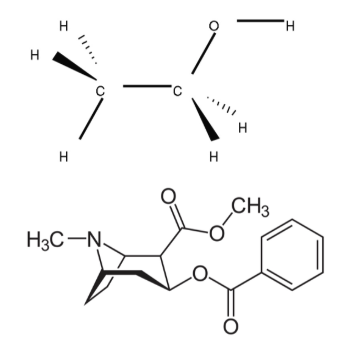
Applying selective pharmacological agents in ex vivo and in vivo preparations allows for assessment of the contribution of receptors and transporters to signaling and behaviors. In addition to using pharmacological agents as tools for probing receptor systems, we also examine the mechanisms of abused compounds to better understand how these drugs affect neurotransmission.
Biphasic Mechanisms of Amphetamine Action at the Dopamine Terminal ➟
Siciliano CA, Calipari ES, Ferris MJ, and Jones SR (2014). Journal of Neuroscience, 34(16):5575-82. doi: 10.1523/JNEUROSCI.4050-13.2014.
α6β2 Subunit Containing Nicotinic Acetylcholine Receptors Exert Opposing Actions on Rapid Dopamine Signaling in the Nucleus Accumbens of Rats With High-Versus Low-Response to Novelty ➟
Siciliano CA, McIntosh JM, Jones SR, and Ferris MJ (2017). Neuropharmacology, 126:281-291. doi: 10.1016/j.neuropharm.2017.06.028.
Optical and electrochemical dopamine sensing
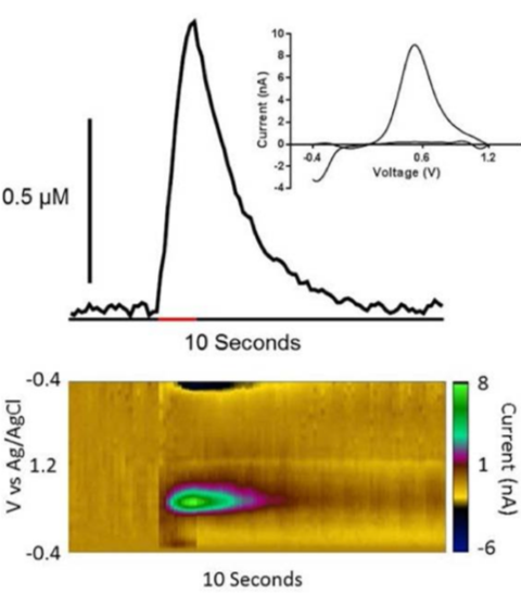
Fast-scan cyclic voltammetry with carbon fiber microelectrodes is an electrochemical method that allows for real-time quantitative assessment of molar concentrations of dopamine within a heterogeneous substance, such as brain tissue. Fluorescent dopamine biosensors provide an alternative way to measure dopamine fluctuations based on changes in fluorescence intensity over time. We leverage both electrochemical and fluorescent sensors for monitoring dopamine dynamics in ex vivo slices and during behavior to interrogate dopamine’s role in decision-making, motivation, and drug-induced plasticity.
Medial prefrontal dopamine dynamics reflect allocation of selective attention ➟Melugin PR, Nolan SO, Kandov E, Ferrara CF, Farahbakhsh ZZ, Siciliano CA (2024). bioRxiv. doi: 10.1101/2024.03.04.583245
Synchrony between midbrain gene transcription and dopamine terminal regulation is modulated by chronic alcohol drinking ➟Farahbakhsh ZZ, Holleran KM, Sens JP, Fordahl SC, Mauterer MI, López AJ, Cuzon Carlson VC, Kiraly DD, Grant KA, Jones SR, Siciliano CA (2024). bioRxiv. doi: 10.1101/2024.03.15.584711.
Neural circuit imaging and manipulation
We use a variety of viral tracing approaches to assess the structure of neural circuits. By injecting viruses carrying fluorescent molecules that move anterograde, retrograde, or transsynaptic, we are able to visualize where brain regions project and from where they receive input. Overlaying this anatomical information with protein and RNA labeling, using immunohistochemistry and in situ hybridization, allows for assessing both the anatomy and molecular identity of cells.
Additionally, recent technological advances have produced a wide array of light-sensitive proteins that can be used to visualize and control specific aspects of cellular activity. Combining optogenetic indicators and actuators allows for “all-optical” circuit dissection: the ability to simultaneously observe and manipulate neuronal activity with cellular resolution and millisecond precision.
We use miniaturized head-mounted microscopes and two-photon microscopy with gradient index lenses which allows us to leverage these approaches for recording and manipulating neuronal activity in the deepest regions of the rodent brain ( below the ventral surface). Our lab is able to chronically record previously inaccessible structures, including the trigeminal ganglion and brainstem nuclei, allowing stable imaging of the same neurons over months in awake, behaving animals.
Nolan SO, Melugin PR, Erickson KR, Adams WR, Farahbakhsh ZZ, Mcgonigle CE, Kwon MH, Costa VD, Hackett TA, Carlson VC, Constantinidis C. Current Biology. 2025 Jan 20;35(2):431-43.
A Cortical-Brainstem Circuit Predicts and Governs Compulsive Alcohol Drinking ➟Siciliano CA, Noamany H, Chang CJ, Brown AR, Chen X, Leible D, Lee JJ, Wang J, Vernon AN, Vander Weele CM, Kimchi EY, Heiman M, and Tye KM (2019). Science. doi: 10.1126/science.aay1186.
Leveraging Calcium Imaging to Illuminate Circuit Dysfunction in Addiction ➟Siciliano CA and Tye KM (2019). Alcohol. doi: 10.1016/j.alcohol.2018.05.013.
Dopamine Enhances Signal-to-Noise Ratio in Cortical-Brainstem Encoding of Aversive Stimuli ➟Vander Weele CM, Siciliano CA, Matthews GA, Namburi P, Izadmehr EM, Espinel IC, Nieh EH, Schut EHS, Padilla-Coreano N, Burgos-Robles A, Chang CJ, Kimchi EY, Beyeler A, Wichmann R, Wildes CP, and Tye KM (2018). Nature. doi: 10.1038/s41586-018-0682-1.